
Inside the tumour
An insight into current research projects

Inside the tumour
An insight into current research projects



Why do some tumours fend off the immune system so effectively? Why does a treatment initially work – and then fail? Four researchers, whose projects are being funded by the Wilhelm Sander Foundation in its anniversary year 2025, provide an insight into their work and demonstrate just how diverse the approaches in cancer research can be today: from molecular transporters in head and neck tumours, through the enigmatic dual nature of a signalling pathway in the brain, to the search for the key to treatment resistance in leukaemia, and on to the mechanisms that turn immune cells in paediatric brain tumours into allies of the cancer.
When the immune system falls silent
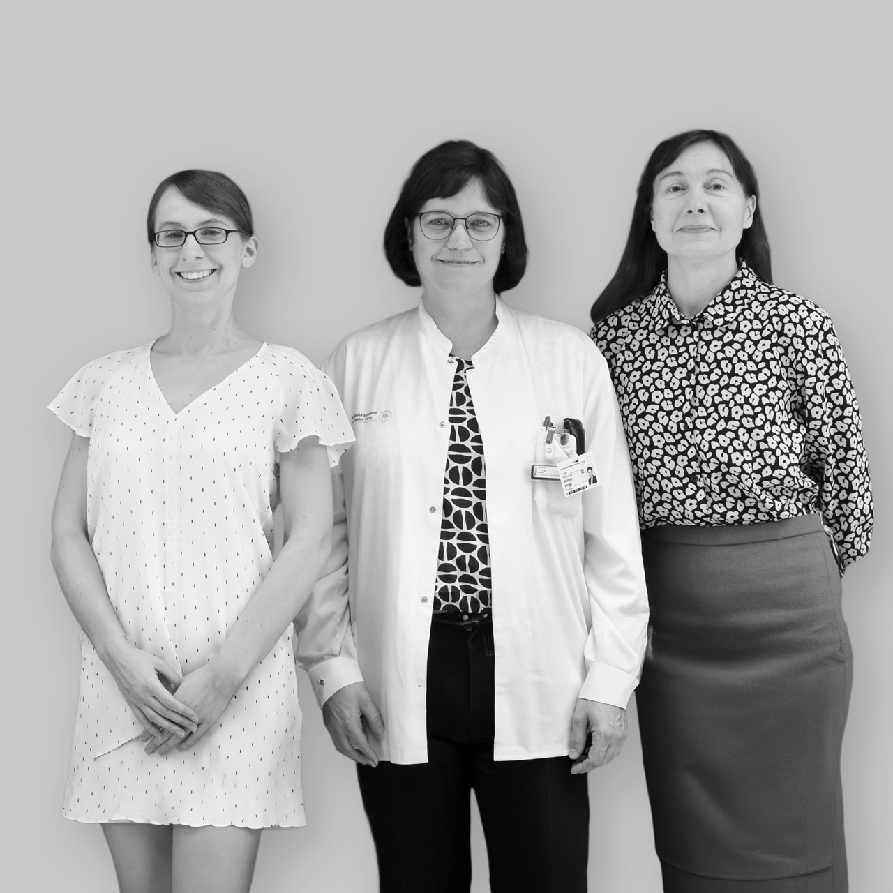
ANNA DUBROVSKA, CLAUDIA ARNDT, ANNETT LINGE, Dresden University of Technology
The starting point was a systematic laboratory comparison: the team led by Anna Dubrovska at Dresden University of Technology investigated membrane proteins from normal tumour cells as well as from those that had been specifically bred to be radiation-resistant. One particular protein kept cropping up: CD98hc is a transporter that channels amino acids – the building blocks of life – through the cell membrane. Clinical data from Annett Linge and her team confirmed the suspicion: tumours in which this protein is particularly active respond less well to chemoradiotherapy and are more likely to recur.
Dubrovska’s research focuses on head and neck squamous cell carcinomas (HNSCC), i.e. tumours that develop, for example, in the throat, on the floor of the mouth or in the larynx. In cases of locally advanced disease, the five-year survival rate is around 65 per cent. HPV-negative cases are particularly difficult to treat. These tumours are resistant to radiotherapy and chemotherapy and skilfully keep the body’s immune system at bay.
Radiotherapy and immunotherapy – better when used together
According to Dubrovska’s hypothesis, CD98hc is not only a marker of resistance but also plays an active role in suppressing the immune response. By reprogramming the metabolism of tumour cells, it alters the tumour’s chemical environment in such a way that immune cells can barely penetrate it or become effective. In three-dimensional tumour models – that is, tissue spheroids grown in the laboratory that mimic a real tumour – the team is investigating whether immune cells can penetrate the tumour tissue more effectively when CD98hc is switched off.
Dubrovska describes the real source of hope as follows: “Our hypothesis is that the combination of radiotherapy and CAR-T cells is more effective than either treatment on its own – particularly for immunocold tumours such as HPV-negative HNSCC.” “CAR-T cells are genetically modified immune cells that can be programmed to target tumour cells specifically,” explains Claudia Arndt. The finding that radiotherapy can further activate the immune system under certain conditions is a promising one – and it is precisely this effect that Dubrovska, Arndt and Linge aim to harness for HNSCC. In parallel, their team is analysing tissue samples from 150 patients to establish CD98hc as a biomarker. In future, this should help predict which patients will benefit most from the combination therapy.
When the immune system falls silent
ANNA DUBROVSKA, CLAUDIA ARNDT, ANNETT LINGE, Dresden University of Technology
The starting point was a systematic laboratory comparison: the team led by Anna Dubrovska at Dresden University of Technology investigated membrane proteins from normal tumour cells as well as from those that had been specifically bred to be radiation-resistant. One particular protein kept standing out: CD98hc is a transporter that channels amino acids – the building blocks of life – through the cell membrane. Clinical data from Annett Linge and her team confirmed the suspicion: tumours in which this protein is particularly active respond less well to chemoradiotherapy and are more likely to recur.
Dubrovska’s research focuses on head and neck squamous cell carcinomas (HNSCC), i.e. tumours that develop, for example, in the throat, on the floor of the mouth or in the larynx. In cases of locally advanced disease, the five-year survival rate is around 65 per cent. HPV-negative cases are particularly difficult to treat. These tumours are resistant to radiotherapy and chemotherapy and skilfully keep the body’s immune system at bay.
Radiotherapy and immunotherapy – better when used together
According to Dubrovska’s hypothesis, CD98hc is not only a marker of resistance but also plays an active role in suppressing the immune response. By reprogramming the metabolism of tumour cells, it alters the tumour’s chemical environment in such a way that immune cells can barely penetrate it or become effective. In three-dimensional tumour models – that is, tissue spheroids grown in the laboratory that mimic a real tumour – the team is investigating whether immune cells can penetrate the tumour tissue more effectively when CD98hc is switched off.
Dubrovska describes the real source of hope as follows: “Our hypothesis is that the combination of radiotherapy and CAR-T cells is more effective than either treatment on its own – particularly for immunocold tumours such as HPV-negative HNSCC.” “CAR-T cells are genetically modified immune cells that can be programmed to target tumour cells specifically,” explains Claudia Arndt. The finding that radiotherapy can further activate the immune system under certain conditions is a promising one – and it is precisely this effect that Dubrovska, Arndt and Linge aim to harness for HNSCC. In parallel, their team is analysing tissue samples from 150 patients to establish CD98hc as a biomarker. In future, this is intended to predict which patients will benefit most from the combination therapy.
A signalling pathway with two sides
CLAUDIO GIACHINO, University of Basel
Tumour cells do not grow in a vacuum. They are embedded in a dense network of neighbouring cells, signalling molecules and immune cells, which they actively exploit for their own benefit. “Brain tumours grow within a complex ecosystem,” says project coordinator Claudio Giachino from the Department of Biomedicine at the University of Basel. Within this ecosystem, tumour cells interact with many other cell types in the surrounding tissue. Gliomas, the most common and aggressive form of brain tumour, are not so difficult to treat simply because the tumour cells themselves are particularly resistant. They skilfully exploit their environment, shut down the immune system and thus create a protective space that therapies can barely penetrate.
Giachino’s research group focuses on the Notch signalling pathway, a highly conserved cell communication system found in almost all multicellular organisms, which determines the role a cell plays within the overall structure. In cancer research, Notch was long regarded as a driver, as the signalling pathway confers stem cell-like properties on tumour cells and makes them more resistant to treatment. Then came the surprising discovery: “We realised that the Notch signalling pathway can inhibit – rather than promote – the growth of certain brain tumour subtypes.” The same signalling pathway, two opposing effects – depending on the tumour type.
Making the tumour vulnerable
This ambivalence opens up new therapeutic possibilities. Recent laboratory findings suggest that increased Notch activity makes tumour cells more sensitive to the immune system, specifically to interferon-gamma, a messenger substance used by immune cells to inhibit and kill cancer cells. To unravel these mechanisms, Giachino’s team is conducting targeted experiments in mouse models and human tumour cell lines. Sometimes Notch activity is increased, sometimes it is switched off – and each time, the team observes how the tumour and its surroundings react. The findings could have implications far beyond gliomas, as Notch signalling pathways and immune cell interactions play a role in many types of cancer.

A signalling pathway with two sides
CLAUDIO GIACHINO, University of Basel
Tumour cells do not grow in a vacuum. They are embedded in a dense network of neighbouring cells, signalling molecules and immune cells, which they actively exploit for their own benefit. “Brain tumours grow within a complex ecosystem,” says project coordinator Claudio Giachino from the Department of Biomedicine at the University of Basel. Within this ecosystem, tumour cells interact with many other cell types in the surrounding tissue. Gliomas, the most common and aggressive form of brain tumour, are not so difficult to treat simply because the tumour cells themselves are particularly resistant. They skilfully exploit their environment, shut down the immune system and thus create a protective space into which therapies can barely penetrate.
Giachino’s research group focuses on the Notch signalling pathway, a highly conserved cell communication system found in almost all multicellular organisms, which determines the role a cell plays within the overall structure. In cancer research, Notch was long regarded as a driver, as the signalling pathway confers stem cell-like properties on tumour cells and makes them more resistant to treatment. Then came the surprising discovery: “We realised that the Notch signalling pathway can inhibit – rather than promote – the growth of certain brain tumour subtypes.” The same signalling pathway, two opposing effects – depending on the tumour type.
Making the tumour vulnerable
This ambivalence opens up new therapeutic possibilities. Recent laboratory findings suggest that increased Notch activity makes tumour cells more sensitive to the immune system, specifically to interferon-gamma, a signalling molecule used by immune cells to inhibit and kill cancer cells. To unravel these mechanisms, Giachino’s team is conducting targeted experiments in mouse models and human tumour cell lines. Sometimes Notch activity is increased, sometimes it is switched off – and each time, the team observes how the tumour and its surroundings react. The findings could have implications far beyond gliomas, as Notch signalling pathways and immune cell interactions play a role in many types of cancer.
When cancer rewrites the rules
CARSTEN MÜLLER-TIDOW, Heidelberg University Hospital
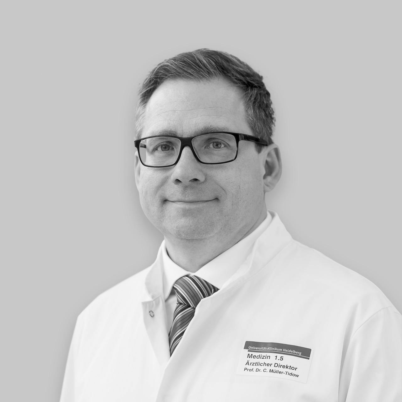
Treatment resistance and relapse remain the greatest clinical challenges in acute myeloid leukaemia (AML). Despite an initial response to treatment, the disease returns in many patients – often in a more resistant form than before. Prof. Carsten Müller-Tidow from Heidelberg University Hospital is investigating the molecular causes of this pattern. His research group has identified a key mechanism in this process: the loss of the EZH2 protein. As an epigenetic regulator, EZH2 determines which genes are active in a cell by influencing the organisation and accessibility of DNA, but not the DNA itself. If this protein is lost, the activation pattern of the cancer cells changes fundamentally, meaning that chemotherapeutic agents that were previously effective no longer work.
From the laboratory to the clinic – and back
The key finding: EZH2 can be restored by inhibiting the proteasome – the cell’s ‘rubbish collection’ system that breaks down excess proteins. If this process is halted, EZH2 levels rise again, and the tumour cells once more become sensitive to chemotherapy. This preclinical finding led to the TEAM trial, a clinical trial in which treatment-resistant AML patients were treated with the proteasome inhibitor bortezomib. The samples from this study now form the basis for the current project: using state-of-the-art single-cell sequencing techniques, Müller-Tidow’s team is analysing which molecular characteristics are associated with response to treatment or resistance, such as DNA mutations, surface proteins or changes in the proteome. The aim is to establish biomarkers that will, in future, predict which patients will benefit from proteasome inhibitor-based therapy. This would represent a step towards a more targeted, personalised treatment for AML.
When cancer rewrites the rules
CARSTEN MÜLLER-TIDOW, Heidelberg University Hospital
Treatment resistance and relapse remain the greatest clinical challenges in acute myeloid leukaemia (AML). Despite an initial response to treatment, the disease returns in many patients – often in a more resistant form than before. Prof. Carsten Müller-Tidow from Heidelberg University Hospital is investigating the molecular causes of this pattern. His research group has identified a key mechanism in this process: the loss of the EZH2 protein. As an epigenetic regulator, EZH2 determines which genes are active in a cell by influencing the packaging and readability of the DNA, but not the DNA itself. If this protein is lost, the activation pattern of the cancer cells changes fundamentally, meaning that chemotherapeutic agents that were previously effective no longer work.
From the laboratory to the clinic – and back
The key finding: EZH2 can be restored by inhibiting the proteasome – the cell’s ‘rubbish collection’ system that breaks down excess proteins. If this process is halted, EZH2 levels rise again, and the tumour cells once more become sensitive to chemotherapy. This preclinical finding led to the TEAM trial, a clinical trial in which treatment-resistant AML patients were treated with the proteasome inhibitor bortezomib. The samples from this study now form the basis for the current project: using state-of-the-art single-cell sequencing techniques, Müller-Tidow’s team is analysing which molecular characteristics are associated with response to treatment or resistance, such as DNA mutations, surface proteins or changes in the proteome. The aim is to establish biomarkers that will, in future, predict which patients will benefit from proteasome inhibitor-based therapy. This would represent a step towards a more targeted, personalised treatment for AML.
When immune cells become tumour helpers
MARIUS MADER, University Medical Centre Hamburg-Eppendorf
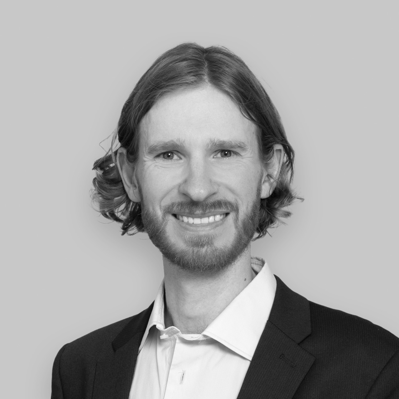
Children with high-grade gliomas – the most aggressive form of brain tumour in childhood – have virtually no chance of recovery despite intensive treatment, as curative treatment options do not yet exist. Dr Marius Mader from the University Medical Centre Hamburg-Eppendorf is researching the pathobiology of this disease, with a particular focus on the tumour microenvironment – that is, the cellular environment in which a tumour grows and which plays a decisive role in determining its characteristics.
As a postdoctoral researcher at Stanford University, Mader carried out pioneering work in the field of microglial replacement. He demonstrated that the brain’s resident immune cells can be systematically replaced by external donor cells. This concept now forms the foundation of his research group, as microglia play a crucial role in brain tumours: they are reprogrammed by tumour cells and thus promote tumour growth rather than fighting it. However, how this reprogramming takes place is still poorly understood, not least because available patient samples from paediatric gliomas are rare and are usually only collected at advanced stages of the disease.
New targets in early tumour stages
The project aims to bridge this knowledge gap. Mader and his team are developing a humanised mouse model into which human microglia derived from induced pluripotent stem cells are introduced and subsequently exposed to paediatric glioma cells. Using this platform, it is possible for the first time to observe the reprogramming of microglia as early as the initial stages of tumour development. This involves recording changes in phenotype and spatial distribution, as well as the signalling pathways between immune cells and tumour cells. This is achieved using single-cell RNA sequencing and spatial transcriptomics. The aim is to identify key mechanisms of pro-tumoural reprogramming and thereby uncover new therapeutic targets for one of the deadliest forms of childhood cancer.
When immune cells become tumour helpers
MARIUS MADER, University Medical Centre Hamburg-Eppendorf
Children with high-grade gliomas – the most aggressive form of brain tumour in childhood – have virtually no chance of recovery despite intensive treatment, as curative treatment options do not yet exist. Dr Marius Mader from the University Medical Centre Hamburg-Eppendorf is researching the pathobiology of this disease, with a particular focus on the tumour microenvironment – that is, the cellular environment in which a tumour grows and which plays a decisive role in determining its characteristics.
As a postdoctoral researcher at Stanford University, Mader carried out pioneering work in the field of microglial exchange. He demonstrated that the brain’s resident immune cells can be systematically replaced by external donor cells. This concept now forms the foundation of his research group, as microglia play a crucial role in brain tumours: they are reprogrammed by tumour cells and thus promote tumour growth rather than fighting it. However, how this reprogramming takes place is still poorly understood, not least because available patient samples from paediatric gliomas are rare and are usually only collected at advanced stages of the disease.
New targets in early tumour stages
The project aims to bridge this knowledge gap. Mader and his team are developing a humanised mouse model into which human microglia derived from induced pluripotent stem cells are introduced and subsequently exposed to paediatric glioma cells. Using this platform, it is possible for the first time to observe the reprogramming of microglia as early as the initial stages of tumour development. This involves recording changes in phenotype and spatial distribution, as well as the signalling pathways between immune cells and tumour cells. This is achieved using single-cell RNA sequencing and spatial transcriptomics. The aim is to identify key mechanisms of pro-tumoural reprogramming and thereby uncover new therapeutic targets for one of the deadliest forms of childhood cancer.